
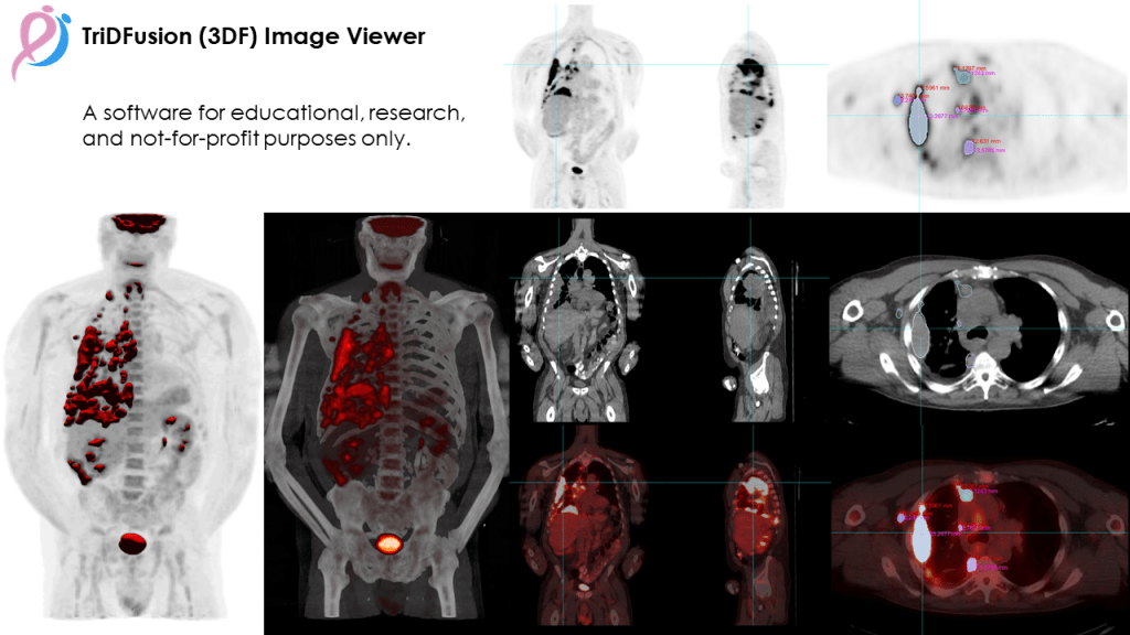
The TriDFusion (3DF) Image Viewer is a MATLABTM-based Multi-Fusion Imaging research software tool designed for physicians, physicist and academic researchers in medical imaging.
It is a Multi-Modality & Multidimensional DICOM Image Viewer with dynamic memory allocation. It can be used to solve issues like image orientation, image slice separation, miss registration and removed and set hot pixels, like the bladder of a Whole-Body SPECT planar, to a new custom value. The mask and image segmentation tool works on any modality. The result image can be exported to a new DICOM series and to a .stl format for 3D printing. The fusion of MIP with isosurface using TriDFusion improves detection and characterization of lesions on 18F-FDG PET/CT compared to evaluation on the MIP only. The addition of volume rendering with customized color and alpha map may provide additional information about the intensity of the uptake in lesions. The 3D threshold base isosurface can be exported as stl and also to a new dicom 3D mask for tumor quantification.
Main features
- Multi-modality Image Viewer
- Total Tumor Burden Determination
- 3D Visualization (3D CT WB, 3D PET AC, 3D small animal, 3D MRI, 2D PET views,
- 3D Printing
- Image Multi-Fusion
- Image Convolution
- Image Registration
- Image Resampling
- Image Re-Orientation
- Image Arithmetic and Post Filtering
- Image Editing
- Contour Segmentation
- Image Mask
- Image Constraint
- Lung Segmentation
- Digital Radiography Lung
- Edge Detection
- Voxel Dosimetry
- Machine Learning Segmentation (supported by the integrated TotalSegmentator tool)
- Radiomics (supported by the PyRadiomics package)
Source Code: Github Page, MATLAB Runtime Library
Documentation: File Format Compatibility, Installation Guide, Usage Guide
Reference Publication (D Lafontaine et al, 2022 EJNMMI Commentary Article)
